Technology

Harnessing this non-classical insight into cellular systems requires quantitative multiscale measurements of coupled excitable dynamics in living systems. We developed an image analysis toolset that reaches from the submicron scale, were optical flow measurements capture pixel scale dynamics, to active contour algorithms for shape dynamics, to particle image velocimetry (PIV) measurements of large scale flow fields in cell groups. Integrating measurements on multiple scales is proving particularly powerful to dissect complex signaling pathways. This systematic quantification toolbox will be the foundation for model development.


Optical Flow Analysis


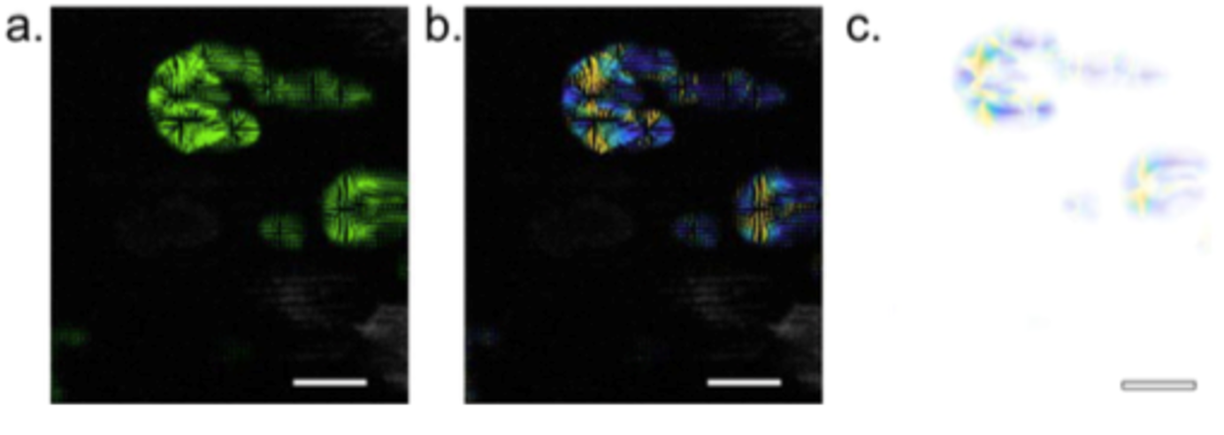
We developed an optical flow analysis algorithm to quantify the speed of cytoskeletal and signaling waves obtained by imaging fluorescent biosensors. Our optical flow analysis uses the Lucas-Kanade algorithm to capture the directionality of cytoskeletal and signaling wave dynamics. After clustering the vectors into single objects, Crocker-Grier's Particle Tracking Algorithm can be used to track the wave fronts.

Optical Flow Analysis

Vector field from optical flow analysis of an HL60 cell on nanotopography with EF cathode to left. (a) Output optical flow vector field. (b) Vector field with vectors color indicating direction. (c) Flow vectors clustered by direction.


Nanotopography Fabrication


Micro- and nano-topographical surfaces are of great interest to probe mechanical perturbations to cell migration and actin dynamics. To fabricate these surfaces, master patterns are designed using multiphoton absorption polymerization (MAP). They can then be replicated using soft lithography and replica molding. We are working on ways to increase the scale of patterns and of output.

Nanotopography and Cell Response

Electron micrograph of ridge nanotopography from Driscoll, Meghan K., et al. "Cellular contact guidance through dynamic sensing of nanotopography." ACS Nano 8.4 (2014): 3546-3555.